What It Is
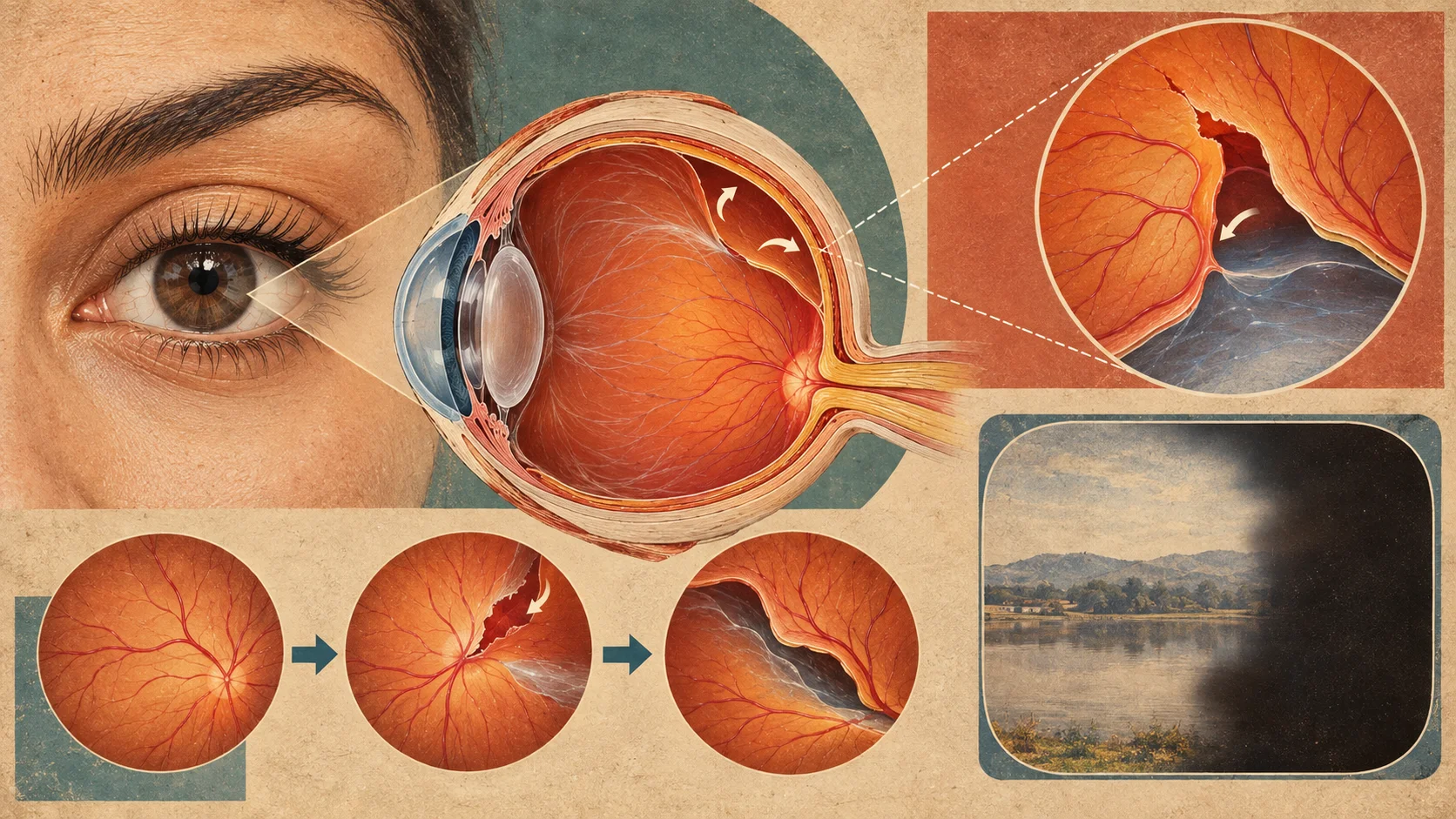
Retinal detachment is a serious condition in which the retina, the light-sensitive layer of tissue at the back of the eye, pulls away from the underlying tissue that supplies it with oxygen and nutrients.
How It Develops
Retinal detachment most commonly occurs when a tear or hole develops in the retina, often related to age-related changes in the gel-like fluid that fills the eye, allowing fluid to seep behind the retina and cause it to separate from the tissue beneath it. It can also occur following significant eye trauma, or in association with certain underlying eye conditions. Once separated from its underlying blood supply, the affected part of the retina can quickly lose function if not treated promptly.
General Symptoms
Common symptoms of retinal detachment include a sudden increase in floaters, flashes of light in the visual field, and a shadow or curtain-like effect spreading across part of the vision. These symptoms typically develop suddenly rather than gradually, which is an important distinguishing feature.
General Treatment Approaches
Retinal detachment is generally considered a medical emergency requiring prompt treatment to preserve vision. Treatment typically involves a surgical procedure aimed at reattaching the retina and sealing any tears, using one of several available surgical techniques depending on the specific characteristics of the detachment.
Background
Retinal detachment was first clearly linked to a tear in the retina in the late 19th century, a discovery that laid the groundwork for developing surgical treatments aimed specifically at repairing that underlying tear. Surgical techniques for retinal reattachment developed considerably over the 20th century, and the condition, once frequently resulting in permanent vision loss, now has a good outcome in many cases when treated promptly after symptoms begin.