What It Is
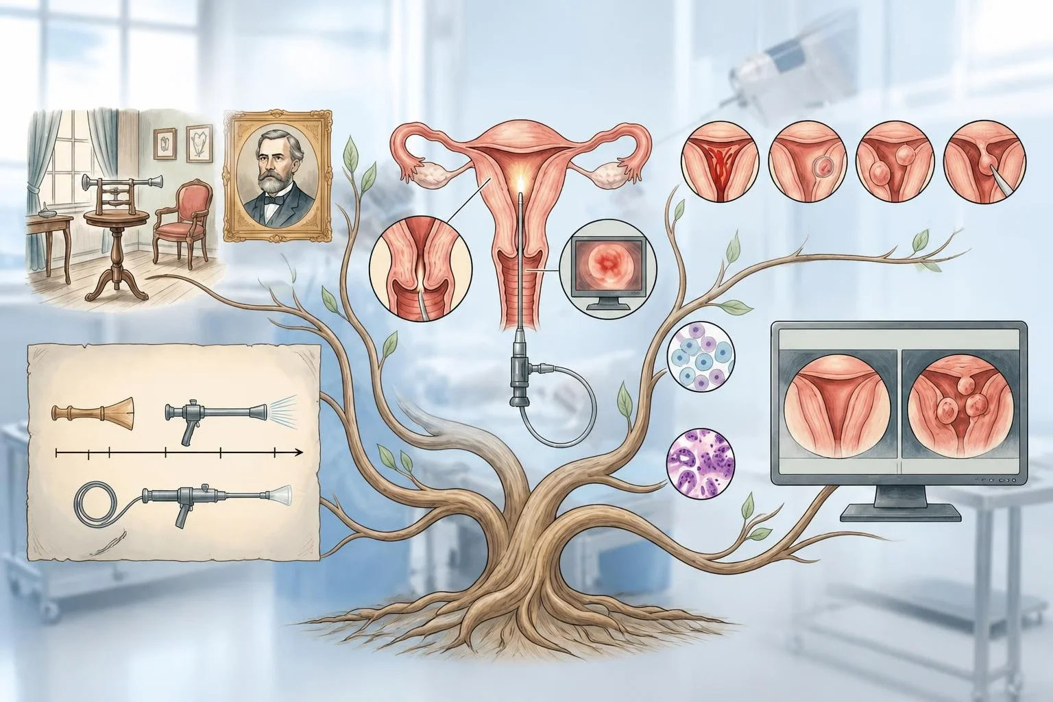
Hysteroscopy is a procedure that allows direct visual examination of the inside of the uterus, using a thin, flexible or rigid tube fitted with a camera, inserted through the cervix.
What It Checks
During a hysteroscopy, the examining physician can directly view the lining of the uterus, looking for abnormalities such as polyps, structural irregularities, or other changes that might not be clearly visible on other imaging tests such as ultrasound.
Why It's Used
Hysteroscopy is commonly used to investigate symptoms such as abnormal uterine bleeding, to evaluate the uterus as part of a fertility assessment, and to perform certain minor procedures, such as removing a polyp, directly during the same session.
How It's Done
During a hysteroscopy, a thin hysteroscope is gently guided through the vagina and cervix into the uterus, often with a small amount of fluid or gas used to gently expand the uterine cavity for a clearer view. The scope transmits real-time video images to a screen for the examining physician to review. The procedure can sometimes be done with minimal or no anesthesia, though sedation is used in some cases depending on the specific circumstances.
Background
Early attempts at examining the inside of the uterus date back to the 19th century, but the technique remained limited by the available instruments and lighting technology of the time. The development of improved fiber-optic and video technology in the latter half of the 20th century transformed hysteroscopy into a much more practical and widely used procedure, and it remains a valuable tool for both diagnosis and minor treatment of conditions affecting the uterine cavity today.