
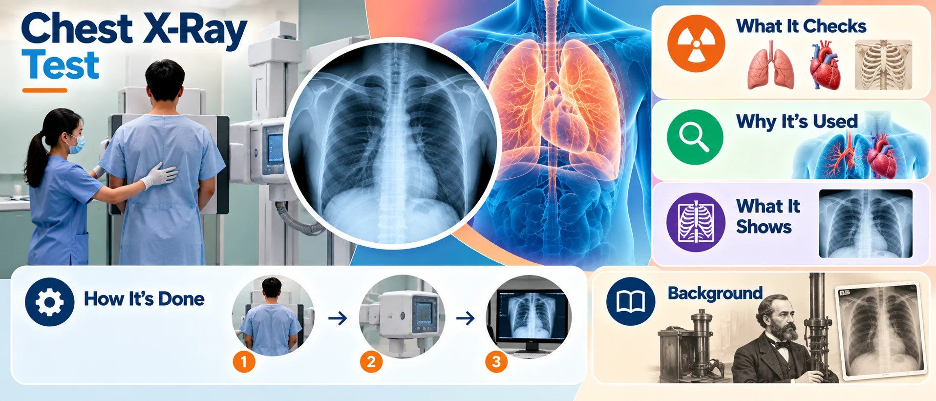
What It Is
A chest X-ray is an imaging test that uses a small amount of radiation to produce a picture of the structures inside the chest, including the lungs, heart, and surrounding bones.
What It Checks
A chest X-ray can show the general size, shape, and position of the lungs and heart, as well as the bones of the chest, including the ribs and spine. Different tissues absorb X-rays to different degrees, which is what allows the resulting image to distinguish between structures like bone, lung tissue, and the heart, showing up as different shades on the final image.
Why It's Used
A chest X-ray is commonly used to evaluate the lungs and heart, investigate symptoms related to breathing or chest discomfort, and as part of general health or pre-procedure evaluations in various clinical settings.
How It's Done
During a chest X-ray, the person typically stands in front of a flat detector plate while an X-ray machine, positioned a short distance away, briefly passes a controlled beam of radiation through the chest. The entire process usually takes only a few minutes, and the resulting image is available for review almost immediately with modern digital equipment.
Background
X-rays were discovered by a German physicist in the late 19th century, a finding that quickly led to their application in medicine, given their ability to reveal internal structures without the need for surgery. Chest X-rays became a standard diagnostic tool in the early 20th century and have remained one of the most commonly performed imaging tests worldwide, valued for being quick, widely accessible, and relatively low in cost compared to more advanced imaging technologies.