What It Is
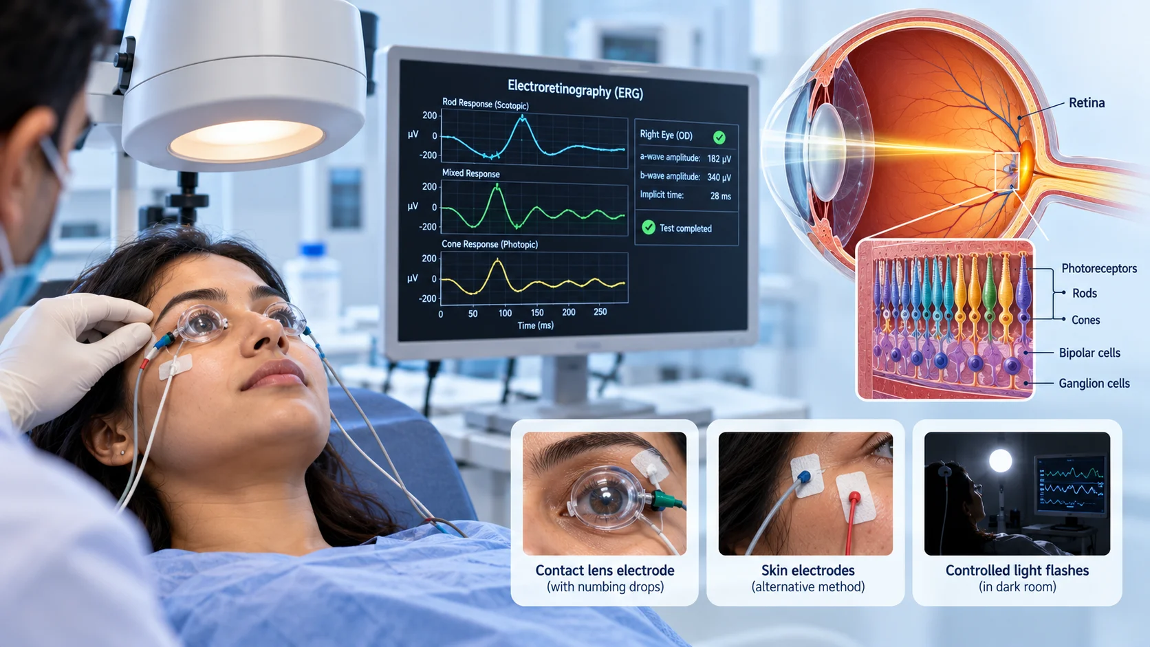
Electroretinography is a test that measures the electrical activity of the retina, the light-sensitive layer of tissue at the back of the eye, in response to light stimulation.
What It Checks
Electroretinography records how the retina's cells respond electrically when exposed to flashes of light, providing information about how well the retina is functioning, including how well its specific light-sensitive cells are working.
Why It's Used
Electroretinography is commonly used to investigate certain retinal conditions, particularly those affecting how the retina responds to light, and can help characterize the specific nature and extent of retinal dysfunction in more detail than a standard eye examination alone might reveal.
How It's Done
During electroretinography, special contact lens electrodes or small skin electrodes are placed on or near the eye, after numbing eye drops are applied for contact-based electrodes. The person is exposed to a series of controlled light flashes, often after a period of adjusting to darkness beforehand, while the resulting electrical activity from the retina is recorded and analyzed.
Background
The retina's electrical response to light was first recorded in the 19th century, but electroretinography developed into a more standardized, clinically useful diagnostic test over the 20th century, aided by advances in electronic recording equipment capable of detecting the retina's relatively small electrical signals. The technique has continued to be refined, and it remains a valuable, specialized tool for evaluating certain retinal conditions in detail today.