What It Is
Magnetic resonance imaging, commonly abbreviated as MRI, is an imaging test that uses strong magnetic fields and radio waves, rather than radiation, to produce detailed images of structures inside the body.
What It Checks
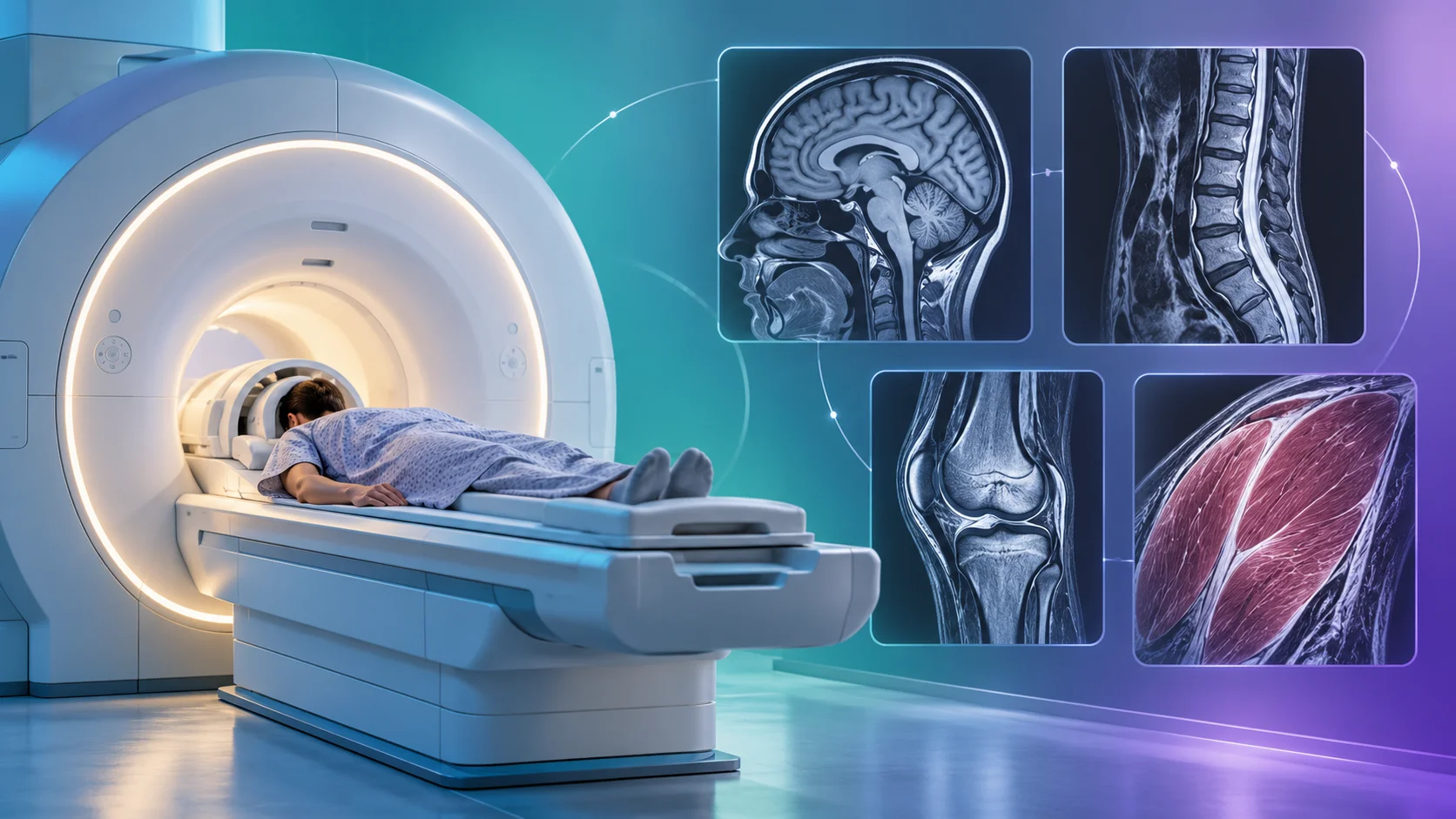
MRI is particularly well suited to producing detailed images of soft tissue, including the brain, spinal cord, muscles, and joints. Depending on the part of the body being examined, an MRI can reveal fine details about tissue structure that aren't as clearly visible using other imaging methods, making it useful for closely examining organs and soft tissue.
Why It's Used
MRI is commonly used to closely examine areas of the body where detailed soft tissue imaging is particularly valuable, including the brain, spine, and joints, and is often used following other initial tests or evaluations when more detailed imaging is needed.
How It's Done
During an MRI, the person lies still inside a large, tube-shaped machine that generates a strong magnetic field. The machine also sends and receives radio wave signals, which interact with water molecules in the body's tissues in a way that allows a computer to construct detailed cross-sectional images. The procedure typically takes anywhere from twenty minutes to an hour, depending on what's being examined, and involves no radiation exposure.
Background
The scientific principles behind MRI, relating to how atomic nuclei respond to magnetic fields, were understood by physicists earlier in the 20th century, but it took until the 1970s and 1980s for researchers to develop these principles into a practical medical imaging technology. Since becoming widely available in the 1980s, MRI has become one of the most valuable tools in modern medical imaging, particularly recognized for its ability to produce detailed images of soft tissue without using radiation.