What It Is
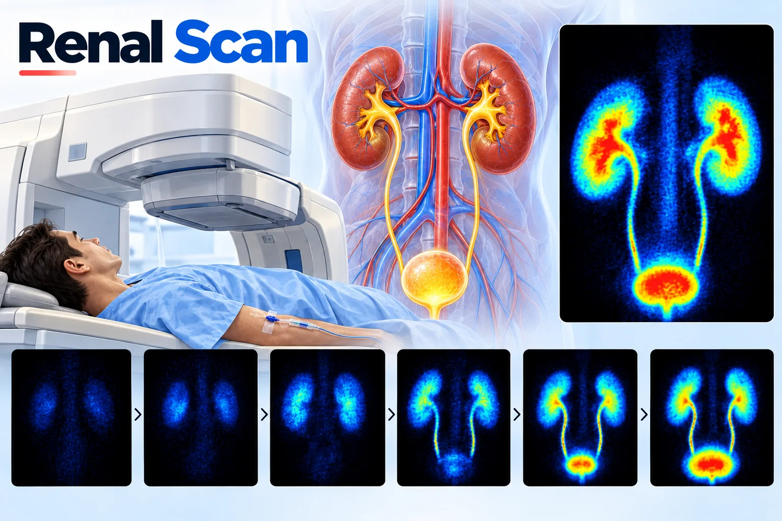
A renal scan is a nuclear medicine imaging test used to evaluate the function of the kidneys, showing how well each kidney is filtering blood and draining urine.
What It Checks
A renal scan uses a small amount of radioactive tracer that the kidneys filter out of the blood, allowing its movement to be tracked as it passes through each kidney and drains into the bladder. This provides information about how well each kidney is functioning individually, including whether blood flow, filtering, and drainage are all proceeding normally.
Why It's Used
A renal scan is commonly used to assess the relative function of each kidney separately, particularly useful in situations where one kidney might be functioning differently from the other, and to investigate suspected blockages affecting urine drainage from a kidney.
How It's Done
Before a renal scan, a small amount of radioactive tracer is injected into a vein, and a special camera then tracks the tracer's movement through the kidneys over a period of time, typically around twenty to thirty minutes, producing images and data that reflect each kidney's individual function.
Background
Renal scanning developed as a nuclear medicine technique in the mid-20th century, growing alongside broader advances in using radioactive tracers to assess organ function directly. It offered a way to evaluate each kidney's individual contribution to overall kidney function, information not easily obtained through blood tests, which reflect the kidneys' combined function, and renal scanning remains a valuable, specialized tool for this specific purpose today.