What It Is
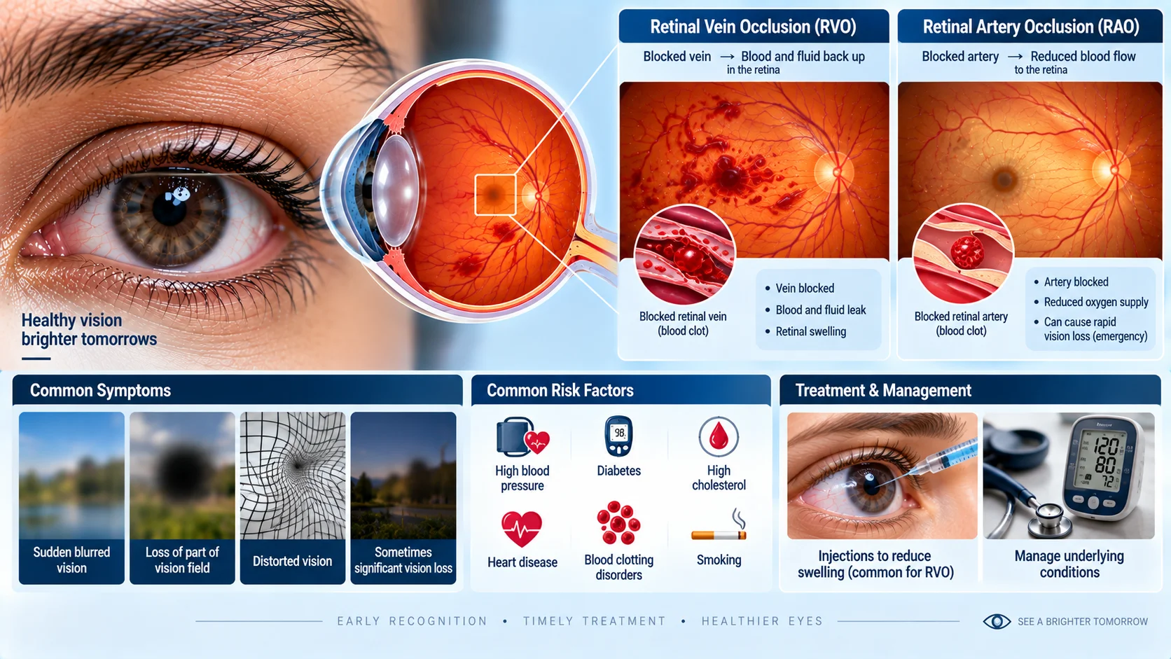
Retinal vein or artery occlusion is a blockage affecting one of the blood vessels supplying the retina, interrupting normal blood flow to part or all of this light-sensitive tissue.
How It Develops
A retinal vein occlusion occurs when a blood clot blocks one of the veins draining blood from the retina, causing blood and fluid to back up within the retinal tissue. A retinal artery occlusion occurs when a clot or other blockage cuts off the blood supply to the retina through one of its arteries, which can rapidly deprive the affected retinal tissue of oxygen. Both types are often related to underlying conditions affecting blood vessels more broadly, such as high blood pressure, diabetes, or conditions affecting blood clotting.
General Symptoms
The main symptom of both types of retinal occlusion is a sudden, usually painless change in vision, which can range from blurring to a significant loss of vision in part or all of the visual field of the affected eye, depending on which vessel is involved and how much of the retina it supplies.
General Treatment Approaches
Retinal artery occlusion is generally considered a medical emergency, since retinal tissue can be permanently damaged within a short time without adequate blood flow, and treatment focuses on trying to restore blood flow as quickly as possible. Retinal vein occlusion is managed somewhat differently, often involving treatments aimed at reducing swelling and complications within the retina, alongside addressing any underlying contributing conditions.
Background
Retinal vein and artery occlusions were recognized as distinct clinical patterns in the 19th century, following the development of the ophthalmoscope, which for the first time allowed direct visual examination of the retina's blood vessels. Understanding of the underlying causes and the development of more targeted treatments for the complications of retinal vein occlusion in particular have advanced considerably in recent decades.