What It Is
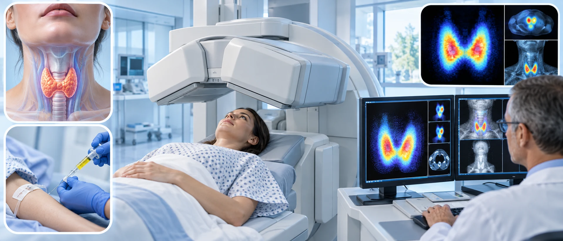
A thyroid scan is a nuclear medicine imaging test that shows how the thyroid gland is functioning, using a small amount of radioactive material that the thyroid naturally absorbs.
What It Checks
Because the thyroid gland naturally absorbs iodine to produce its hormones, a thyroid scan uses a small amount of a radioactive tracer, often a form of iodine or a related substance, which the thyroid takes up similarly. The resulting images show which areas of the thyroid are more or less active than expected, helping reveal patterns relevant to various thyroid conditions.
Why It's Used
A thyroid scan is commonly used to investigate certain thyroid abnormalities, such as unusual nodules found on the thyroid, helping determine whether a specific area is functioning more or less actively than the surrounding tissue, which can provide useful diagnostic information.
How It's Done
Before a thyroid scan, a small amount of radioactive tracer is given, either by mouth or by injection, and the person waits a period of time to allow the thyroid to take up the tracer. A special camera is then used to detect the tracer's activity within the thyroid, producing images that show the pattern of activity across the gland.
Background
Nuclear medicine imaging of the thyroid developed in the mid-20th century, following the discovery that the thyroid's natural affinity for iodine could be used diagnostically by substituting a radioactive form of the element. The technique became an established diagnostic tool for evaluating thyroid nodules and certain other thyroid conditions, and it remains a useful, specialized imaging option within the broader range of thyroid-related tests available today.