What It Is
A VQ scan, short for ventilation-perfusion scan, is a nuclear medicine imaging test that evaluates both airflow and blood flow within the lungs.
What It Checks
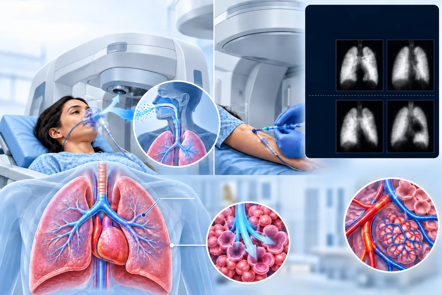
A VQ scan has two parts. The ventilation portion assesses how well air is reaching different areas of the lungs. The perfusion portion assesses how well blood is flowing through the lungs' blood vessels. Comparing these two sets of images together can reveal areas where blood flow and airflow don't match up as expected, a pattern relevant to certain lung conditions.
Why It's Used
A VQ scan is commonly used to investigate a suspected blood clot in the lungs, known as a pulmonary embolism, particularly in situations where other imaging tests, such as CT, aren't suitable for a particular patient.
How It's Done
For the ventilation portion, the person breathes in a small amount of radioactive gas or aerosol, and a special camera captures images showing how the gas distributes throughout the lungs. For the perfusion portion, a small amount of radioactive tracer is injected into a vein, and images are taken showing how blood flows through the lungs' blood vessels. The two sets of images are then compared.
Background
Nuclear medicine techniques for assessing lung ventilation and blood flow developed separately in the mid-20th century before being combined into the standard two-part VQ scan protocol used today. The technique became a widely used method for investigating suspected pulmonary embolism, and while other imaging methods have become more commonly used as a first choice in many settings, VQ scanning remains a valuable diagnostic option, particularly for patients who aren't good candidates for those alternative tests.